Copyright
©2007 Baishideng Publishing Group Co.
World J Gastroenterol. Jun 28, 2007; 13(24): 3384-3387
Published online Jun 28, 2007. doi: 10.3748/wjg.v13.i24.3384
Published online Jun 28, 2007. doi: 10.3748/wjg.v13.i24.3384
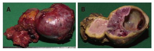
Figure 1 Adrenalectomy specimen from the left side containing a sharply circumscribed tumor (pheochromocytoma) of a x b cm in diameter.
A: The intact resectate; B: A cross section with partial cystic transformation on the right and residual adrenal cortex at the periphery of the tumor.
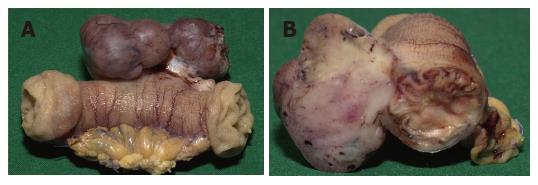
Figure 2 Segmental resectate of the small bowel with a lobulated subserosal tumor mass originating from the muscular bowel wall.
A: The intact resection specimen; B: A cross section perpendicular to the bowel axis.
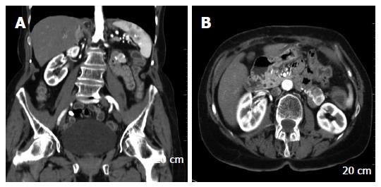
Figure 3 Computed tomography showing the right sided pheochromocytoma (A) and the jejunal GIST (A and B).
- Citation: Kramer K, Hasel C, Aschoff AJ, Henne-Bruns D, Wuerl P. Multiple gastrointestinal stromal tumors and bilateral pheochromocytoma in neurofibromatosis. World J Gastroenterol 2007; 13(24): 3384-3387
- URL: https://www.wjgnet.com/1007-9327/full/v13/i24/3384.htm
- DOI: https://dx.doi.org/10.3748/wjg.v13.i24.3384


