Copyright
©2006 Baishideng Publishing Group Co.
World J Gastroenterol. Sep 28, 2006; 12(36): 5913-5915
Published online Sep 28, 2006. doi: 10.3748/wjg.v12.i36.5913
Published online Sep 28, 2006. doi: 10.3748/wjg.v12.i36.5913
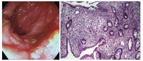
Figure 1 Endoscopy (A) and histological examination (B) showing pouchitis in the ileal J-pouch.
Figure 2 Upper gastrointestinal examination (A), endoscopy (B) and histological examination (C) showing lesions in the stomach.
Figure 3 Hypotonic duodenography (A), endscopy (B) and histological examination (C) showing lesions in the duodenum.
- Citation: Ikeuchi H, Hori K, Nishigami T, Nakano H, Uchino M, Nakamura M, Kaibe N, Noda M, Yanagi H, Yamamura T. Diffuse gastroduodenitis and pouchitis associated with ulcerative colitis. World J Gastroenterol 2006; 12(36): 5913-5915
- URL: https://www.wjgnet.com/1007-9327/full/v12/i36/5913.htm
- DOI: https://dx.doi.org/10.3748/wjg.v12.i36.5913


