Copyright
©2006 Baishideng Publishing Group Co.
World J Gastroenterol. Jan 14, 2006; 12(2): 336-339
Published online Jan 14, 2006. doi: 10.3748/wjg.v12.i2.336
Published online Jan 14, 2006. doi: 10.3748/wjg.v12.i2.336
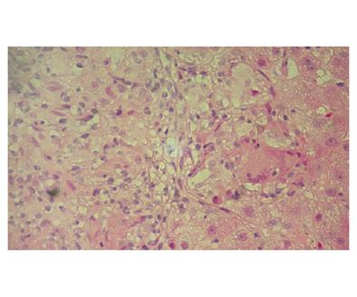
Figure 1 Liver histopathology shows an epithelioid granuloma without central necrosis.
The granuloma is consisted of epithelioid macrophages and some chronic inflammatory cells. There is also a multinucleated giant cell (Hematoxylin and eosin staining x 40).
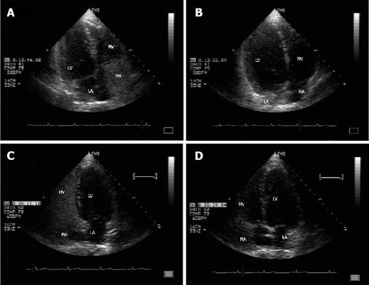
Figure 2 Contrast-enhanced echocardiography image of the patient before (A, B) and after (C, D) corticosteroid treatment for granulomatous hepatitis.
A, C: Microbubble appearance in the right heart chamber (RA and RV) after the bolus infusion of agitated saline; B: Microbubble opacification in the left heart chambers (LA and LV) is not detected after six to eight heart beats; D: Microbubble opacification in the left heart chambers (LA and LV)is not detected after six to eight heart beats.
- Citation: Tzovaras N, Stefos A, Georgiadou SP, Gatselis N, Papadamou G, Rigopoulou E, Ioannou M, Skoularigis I, Dalekos GN. Reversion of severe hepatopulmonary syndrome in a non cirrhotic patient after corticosteroid treatment for granulomatous hepatitis: A case report and review of the literature. World J Gastroenterol 2006; 12(2): 336-339
- URL: https://www.wjgnet.com/1007-9327/full/v12/i2/336.htm
- DOI: https://dx.doi.org/10.3748/wjg.v12.i2.336


