Copyright
©2006 Baishideng Publishing Group Co.
World J Gastroenterol. Jan 14, 2006; 12(2): 246-250
Published online Jan 14, 2006. doi: 10.3748/wjg.v12.i2.246
Published online Jan 14, 2006. doi: 10.3748/wjg.v12.i2.246
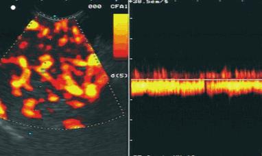
Figure 1 Endoscopic ultrasound image of chronic panreatitis.
Regular vascularisation is shown with detection of venous vessels using contrast-enhanced power Doppler scanning in combination with power Doppler.
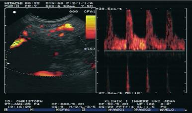
Figure 2 Endoscopic ultrasound image of ductal adenocarcinoma of the pancreas.
Irregular vascularisation is shown with detection of only arterial and no venous vessels using contrast-enhanced power Doppler scanning in combination with pw-Doppler.
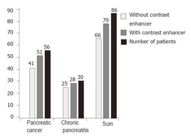
Figure 3 Results of contrast-enhanced endoscopic ultrasound in differentiation between pancreatic carcinoma and focal pancreatitis.
- Citation: Hocke M, Schulze E, Gottschalk P, Topalidis T, Dietrich CF. Contrast-enhanced endoscopic ultrasound in discrimination between focal pancreatitis and pancreatic cancer. World J Gastroenterol 2006; 12(2): 246-250
- URL: https://www.wjgnet.com/1007-9327/full/v12/i2/246.htm
- DOI: https://dx.doi.org/10.3748/wjg.v12.i2.246


