Copyright
©2006 Baishideng Publishing Group Co.
World J Gastroenterol. May 14, 2006; 12(18): 2941-2944
Published online May 14, 2006. doi: 10.3748/wjg.v12.i18.2941
Published online May 14, 2006. doi: 10.3748/wjg.v12.i18.2941
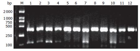
Figure 1 Comparison of the results of RT-PCR and H-E staining.
M: DL 2 000 Marker; Lanes1-12: dissected lymph nodes. Lymph nodes of Lane 7, Lane 10, and Lane 11 were diagnosed lymph node micrometastasis.
Figure 2 Expression of MMP-2 in gastric carcinoma.
A: poorly-differentiated gastric carcinoma (× 400); B: moderately -differentiated gastric carcinoma (× 400); C: well-differentiated gastric carcinoma (× 400).
- Citation: Wu ZY, Li JH, Zhan WH, He YL. Lymph node micrometastasis and its correlation with MMP-2 expression in gastric carcinoma. World J Gastroenterol 2006; 12(18): 2941-2944
- URL: https://www.wjgnet.com/1007-9327/full/v12/i18/2941.htm
- DOI: https://dx.doi.org/10.3748/wjg.v12.i18.2941


