Copyright
©2006 Baishideng Publishing Group Co.
World J Gastroenterol. May 14, 2006; 12(18): 2806-2817
Published online May 14, 2006. doi: 10.3748/wjg.v12.i18.2806
Published online May 14, 2006. doi: 10.3748/wjg.v12.i18.2806
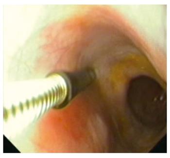
Figure 1 The stimulation electrode visualized in the distal esophagus using the nasal endoscope.
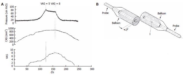
Figure 2 Top corner: The impedance planimetric probe for mechanical stimulation of the gastrointestinal tract.
I and d denote infusion side for filling the balloon and the distance between two detection electrodes. Left: Recordings in the small intestine using the impedance planimetric probe with pressure, cross-sectional area (CSA) and VAS response as function of time during a ramp distension of the balloon up to the pain threshold where the bag volume is kept constant. It appears that the pressure decreased, the CSA was constant whereas the pain continued to increase.
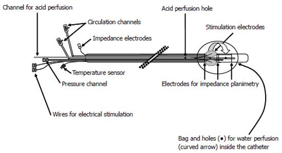
Figure 3 Schematic illustration of the probe used for multi-modal (electrical, mechanical, cold and warmth stimuli) of the esophagus.
The stimulations can be combined with sensitization using acid perfusion of the distal esophagus.
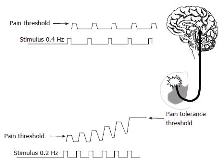
Figure 4 Central integration of repeated stimulation is seen when the stimulation frequency exceeds 0.
3 Hz (bottom picture), whereas slower stimulation does not change the central gain (top picture).
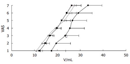
Figure 5 Stimulus-response function (mean ± SEM) to mechanical stimulation in patients with non-erosive gastro-esophageal reflux disease (NERD).
The controls (■, n = 15) and patients with a normal 24-h pH-profile (▲, n = 7) had similar response to the mechanical stimulation, whereas patients with pathological pH-profile (◊, n = 6) showed hypoalgesia to the stimulations. The x-axis shows the volume of the balloon in the distal esophagus and the y-axis denotes the pain response on a visual analogue scale (VAS) with 5 as the pain threshold.
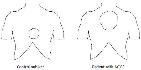
Figure 6 The localization and size of the referred pain area to mechanical stimulations of the distal esophagus in a typical control subject and a patient with non-cardiac chest pain (NCCP).
The patient had and increased size and abnormal localization of the referred pain.
- Citation: Drewes AM, Arendt-Nielsen L, Funch-Jensen P, Gregersen H. Experimental human pain models in gastro-esophageal reflux disease and unexplained chest pain. World J Gastroenterol 2006; 12(18): 2806-2817
- URL: https://www.wjgnet.com/1007-9327/full/v12/i18/2806.htm
- DOI: https://dx.doi.org/10.3748/wjg.v12.i18.2806


