Copyright
©2006 Baishideng Publishing Group Co.
World J Gastroenterol. Jan 7, 2006; 12(1): 94-98
Published online Jan 7, 2006. doi: 10.3748/wjg.v12.i1.94
Published online Jan 7, 2006. doi: 10.3748/wjg.v12.i1.94
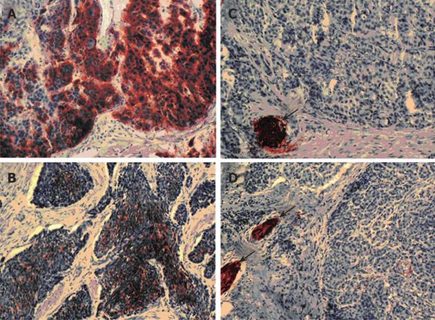
Figure 1 L1 expression in pancreatic neuroendocrine tumours or carcinomas.
Immunohistochemical staining was performed by peroxidase method using monoclonal antibody UJ.127 against L1. Poorly-differentiated L1-positive pancreatic neuroendocrine carcinomas (grade 2; A and B) were shown in comparison to well-differentiated L1-negative tumours (grade 1a; C and D). Peripheral nerves (arrows) stained in (C, D) served as internal positive controls (Magnification ×200 (A and C) and ×400 (B and C)).
- Citation: Kaifi JT, Zinnkann U, Yekebas EF, Schurr PG, Reichelt U, Wachowiak R, Fiegel HC, Petri S, Schachner M, Izbicki JR. L1 is a potential marker for poorly-differentiated pancreatic neuroendocrine carcinoma. World J Gastroenterol 2006; 12(1): 94-98
- URL: https://www.wjgnet.com/1007-9327/full/v12/i1/94.htm
- DOI: https://dx.doi.org/10.3748/wjg.v12.i1.94


