Copyright
©2005 Baishideng Publishing Group Inc.
World J Gastroenterol. Nov 7, 2005; 11(41): 6440-6444
Published online Nov 7, 2005. doi: 10.3748/wjg.v11.i41.6440
Published online Nov 7, 2005. doi: 10.3748/wjg.v11.i41.6440
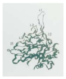
Figure 1 Three-dimensional structure of FHV capsid protein.
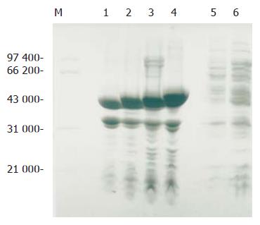
Figure 2 Recombinant proteins expressed in E.
coli cells. M: protein molecular mass standard; lane 1: recombinant vector protein Wt; lane 2: chimeric protein I3S; lane 3: L3C1-I2E-L1C2-L2E; lane 4: L3C1-I2E-L1C2-L2E-I3S; lane 5: supernatant of BL21 cells transformed by pET-L3C1-I2E-L1C2-L2E; lane 6: non-transformed BL21 cells.
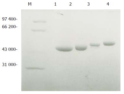
Figure 3 Expression of purified expressed proteins.
M: protein molecular mass standard; lane 1: recombinant vector protein Wt; lane 2: chimeric protein I3S; lane 3: L3C1-I2E-L1C2-L2E; lane 4: L3C1-I2E-L1C2-L2E-I3S.
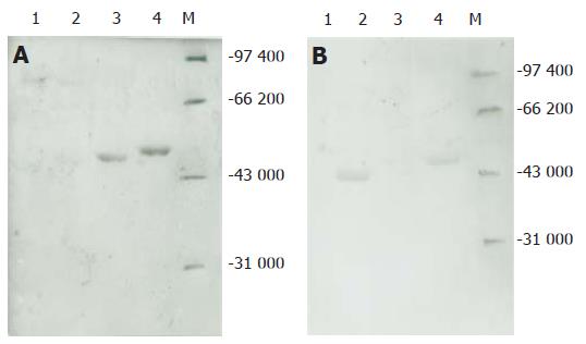
Figure 4 Western blot of expressed proteins using anti-HCV+ (A) and anti-HBsAg+ (B) sera as detecting antibodies.
M: protein molecular mass standard; lane 1: recombinant vector protein W; lane 2: chimeric protein I3S, lane 3: L3C1-I2E-L1C2-L2E; lane 4: L3C1-I2E-L1C2-L2E-I3S.
- Citation: Xiong XY, Liu X, Chen YD. Expression and immunoreactivity of HCV/HBV epitopes. World J Gastroenterol 2005; 11(41): 6440-6444
- URL: https://www.wjgnet.com/1007-9327/full/v11/i41/6440.htm
- DOI: https://dx.doi.org/10.3748/wjg.v11.i41.6440


