Copyright
©The Author(s) 2005.
World J Gastroenterol. Jul 7, 2005; 11(25): 3830-3833
Published online Jul 7, 2005. doi: 10.3748/wjg.v11.i25.3830
Published online Jul 7, 2005. doi: 10.3748/wjg.v11.i25.3830
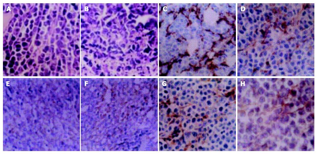
Figure 1 HE staining: morphology of tumor cells in AG group (A, original magnification x100) and control group (B, original magnification x200); immunohistochemical staining of tumor cells: the expression of FVIIIRag (C), iNOS (E) and VEGF (G) in control group, and the expression of FVIIIRag (D), iNOS (F) and VEGF (H) in AG group.
(original magnification, x400).
- Citation: Wang GY, Ji B, Wang X, Gu JH. Anti-cancer effect of iNOS inhibitor and its correlation with angiogenesis in gastric cancer. World J Gastroenterol 2005; 11(25): 3830-3833
- URL: https://www.wjgnet.com/1007-9327/full/v11/i25/3830.htm
- DOI: https://dx.doi.org/10.3748/wjg.v11.i25.3830


