Copyright
©2005 Baishideng Publishing Group Inc.
World J Gastroenterol. Apr 7, 2005; 11(13): 2035-2038
Published online Apr 7, 2005. doi: 10.3748/wjg.v11.i13.2035
Published online Apr 7, 2005. doi: 10.3748/wjg.v11.i13.2035
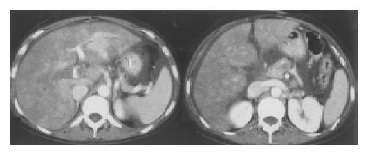
Figure 1 Partial occlusion of the main portal vein (left) and complete occlusion of prossimal superior mesenteric vein (right) shown on abdominal CT-angiogram.
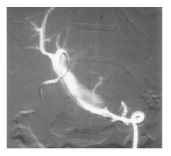
Figure 2 Complete occlusion of the right branch and partial occlusion of the main portal vein and proximal portion of superior mesenteric vein shown by angiography via transhepatic cathether.
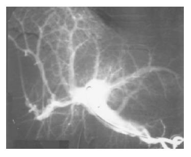
Figure 3 Complete resolution of superior mesenteric and main portal vein thrombosis and partial resolution of thrombi in right portal branch shown by angiography via transhepatic cathether (48 h follow up).
- Citation: Guglielmi A, Fior F, Halmos O, Veraldi GF, Rossaro L, Ruzzenente A, Cordiano C. Transhepatic fibrinolysis of mesenteric and portal vein thrombosis in a patient with ulcerative colitis: A case report. World J Gastroenterol 2005; 11(13): 2035-2038
- URL: https://www.wjgnet.com/1007-9327/full/v11/i13/2035.htm
- DOI: https://dx.doi.org/10.3748/wjg.v11.i13.2035


