Copyright
©2005 Baishideng Publishing Group Inc.
World J Gastroenterol. Jan 7, 2005; 11(1): 61-68
Published online Jan 7, 2005. doi: 10.3748/wjg.v11.i1.61
Published online Jan 7, 2005. doi: 10.3748/wjg.v11.i1.61
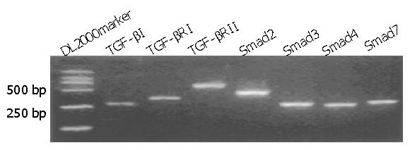
Figure 1 RT-PCR data from BGC-823 cell line demonstrate complete expression of all main components of TGF-β1/Smad signal pathway, including TGF-β1 (284 bp), TβRI (358 bp), TβRII (688 bp), Smad2 (489 bp), Smad3 (284 bp), Smad4 (264 bp) and Smad7 (294 bp).
Products were electrophoresed on 1.5% agarose gel containing ethidium bromide.
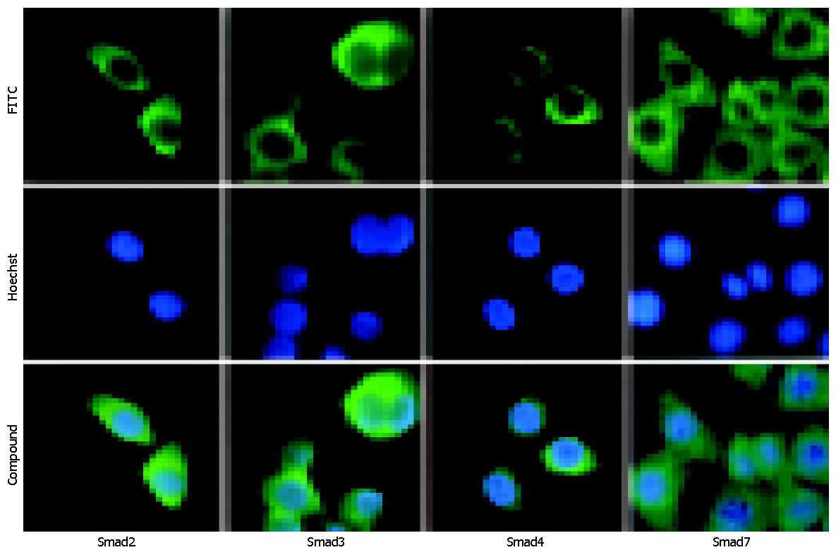
Figure 2 Localization and expression of Smads in BGC-823 cells by immunofluorescence.
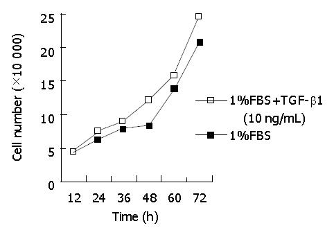
Figure 3 Effect of TGF-β1 on proliferation of BGC-823 cells.
Figure 4 FCM analysis of BGC-823 cells treated with TGF-β1.
A: Controls; B: Treated with TGF-β1 for 24 h; C: Treated with TGF-β1 for 48 h.
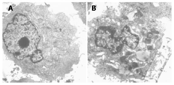
Figure 5 TGF-β1-induced ultrastructural changes in BGC-823 cells under TEM.
A: Controls, ×5000; B: Treated with TGF-β1 for 48 h, ×8000.
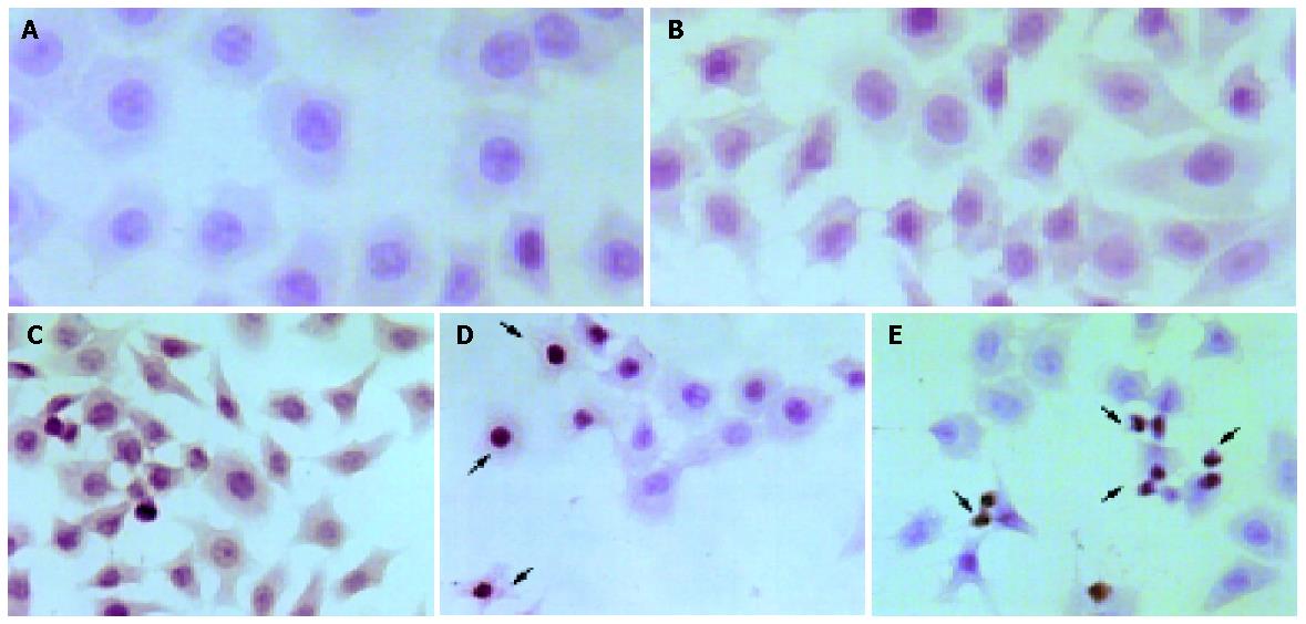
Figure 6 Apoptotic cells induced by TGF-β1 in BGC-823 cells with TUNEL assay (×200).
A: Normal BGC-823 cells; B: Negative controls; C: Positive controls; D and E: Treated with TGF-β1 for 48 h. The arrows in picture D and E are pointed to positive cells.
Figure 7 Up-regulation of endogenous genes in BGC-823 cells treated with TGF-β1.
RT-PCR products were electrophoresed on 1.5% agarose gel containing ethidium bromide. The level of β-actin was used as an internal control. M: DL2000 marker; N: the basal level; lanes 1, 2 and 3: 1-, 2- and 3-h exposure to TGF-β1, respectively.
Figure 8 Increase of p15, p21 and Smad7 proteins confirmed by Western blot analysis.
N: basal level; lanes 1, 2 and 3: 1-, 2- and 3-exposure to TGF-β1, respectively.
- Citation: Li X, Zhang YY, Wang Q, Fu SB. Association between endogenous gene expression and growth regulation induced by TGF-β1 in human gastric cancer cells. World J Gastroenterol 2005; 11(1): 61-68
- URL: https://www.wjgnet.com/1007-9327/full/v11/i1/61.htm
- DOI: https://dx.doi.org/10.3748/wjg.v11.i1.61


