Copyright
©The Author(s) 2004.
World J Gastroenterol. Dec 15, 2004; 10(24): 3616-3620
Published online Dec 15, 2004. doi: 10.3748/wjg.v10.i24.3616
Published online Dec 15, 2004. doi: 10.3748/wjg.v10.i24.3616
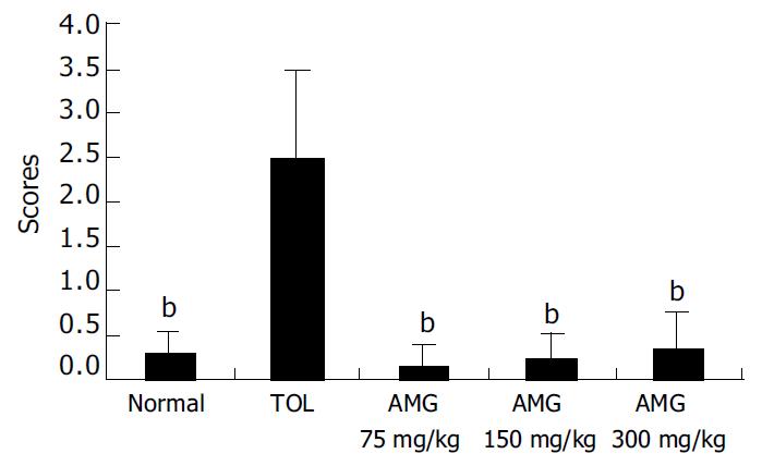
Figure 1 Repeated treatment with AMG on gastric mucosal damage in normal mice (n = 10).
bP < 0.01 vs TOL.
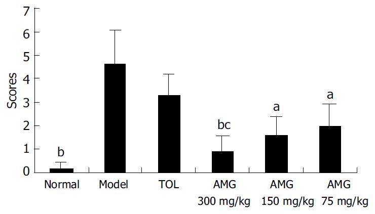
Figure 2 Effect of AMG on ethanol-induced gastric mucosal damage model (n = 10).
aP < 0.05, bP < 0.01 vs model; cP < 0.05, AMG (300 mg/kg) vs TOL.
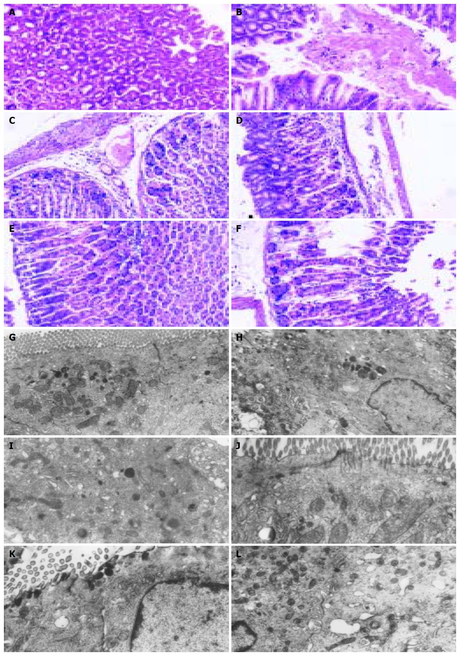
Figure 3 Light and electronic microscopy assessments of ethanol-induced gastric lesion.
A: light microsocopy assessment of ethanol-induced gastric lesion (*100). A: normal group, B: model group, C: ToL group, D: AMG (300 mg/kg) group E: AMG (150 mg/kg) group, F: AMG (75 mg/kg) group. B: Electronic microscopy assessment of ethanol-induced gastric lesion (× 6000) G: normal group, H: model group, I: TOL group, J: AMG (300 mg/kg) group, K: AMG (150 mg/kg) group, L: AMG (75 mg/kg) group.
- Citation: Li YH, Li J, Huang Y, Lü XW, Jin Y. Gastroprotective effect and mechanism of amtolmetin guacyl in mice. World J Gastroenterol 2004; 10(24): 3616-3620
- URL: https://www.wjgnet.com/1007-9327/full/v10/i24/3616.htm
- DOI: https://dx.doi.org/10.3748/wjg.v10.i24.3616


