Copyright
©The Author(s) 2004.
World J Gastroenterol. Nov 1, 2004; 10(21): 3103-3106
Published online Nov 1, 2004. doi: 10.3748/wjg.v10.i21.3103
Published online Nov 1, 2004. doi: 10.3748/wjg.v10.i21.3103
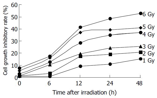
Figure 1 Effect of irradiation on proliferation of HepG2 cells.
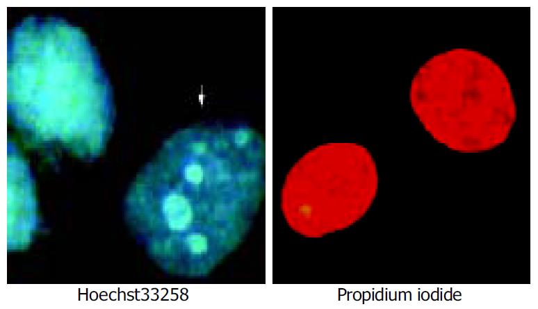
Figure 2 Counterstaining of Hoechst 33258 and propidium iodide to distinguish apoptotic from necrotic cells.
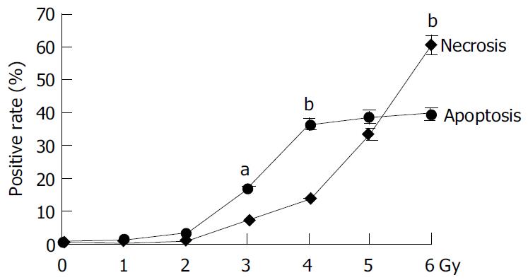
Figure 3 Comparsion of apoptosis and necrosis rate of HepG2 treated by 60Co γ-irradiation.
aP < 0.05, bP < 0.01 vs necrosis.
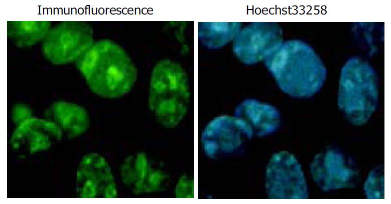
Figure 4 Subcellular localization and expression of p27kip1 in tranfected HepG2 cells.
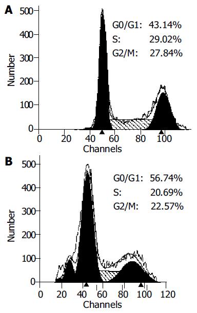
Figure 5 Flow cytometry analysis of cell cycle of nontransfected and transiently transfected HepG2 cells.
A: Nontransfected HepG2 cells. B: p27kip1 transiently transfected HepG2 cells.
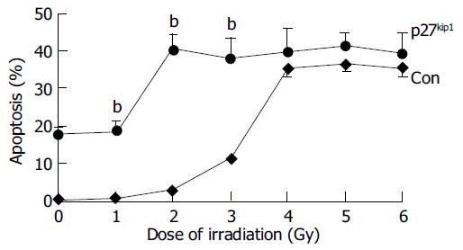
Figure 6 Apoptotic rates induced by γ-irradiation in nontransfected and transfected HepG2 cells.
bP < 0.01 vs control.
- Citation: Guan XX, Chen LB, Ding GX, De W, Zhang AH. Transfection of p27kip1 enhances radiosensitivity induced by 60Co γ-irradiation in hepatocellular carcinoma HepG2 cell line. World J Gastroenterol 2004; 10(21): 3103-3106
- URL: https://www.wjgnet.com/1007-9327/full/v10/i21/3103.htm
- DOI: https://dx.doi.org/10.3748/wjg.v10.i21.3103


