Copyright
©The Author(s) 2004.
World J Gastroenterol. Sep 1, 2004; 10(17): 2598-2601
Published online Sep 1, 2004. doi: 10.3748/wjg.v10.i17.2598
Published online Sep 1, 2004. doi: 10.3748/wjg.v10.i17.2598
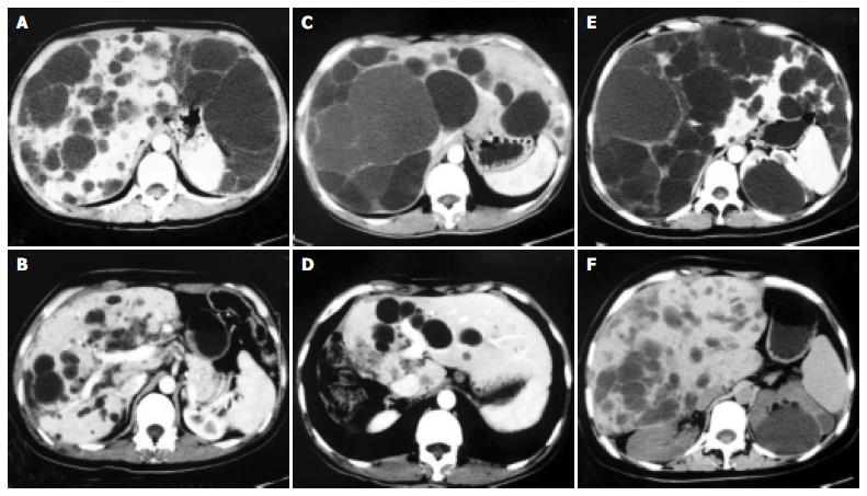
Figure 1 CT scans of livers in PLD patients before (left panels) and 3 mo (right panels) after operation, show that hypertrophy of spared liver and lack of clinically significant cyst progression, and that operative methods totally depend on cystic distribution in different patients, e.
g., lateral segment removal from A to B, right semi-hepatectomy resection from C to D, whereas extensive fenestration of posterior and interior cysts from E to F.
- Citation: Yang GS, Li QG, Lu JH, Yang N, Zhang HB, Zhou XP. Combined hepatic resection with fenestration for highly symptomatic polycystic liver disease: A report on seven patients. World J Gastroenterol 2004; 10(17): 2598-2601
- URL: https://www.wjgnet.com/1007-9327/full/v10/i17/2598.htm
- DOI: https://dx.doi.org/10.3748/wjg.v10.i17.2598


