Copyright
©The Author(s) 2004.
World J Gastroenterol. Jul 1, 2004; 10(13): 1979-1983
Published online Jul 1, 2004. doi: 10.3748/wjg.v10.i13.1979
Published online Jul 1, 2004. doi: 10.3748/wjg.v10.i13.1979
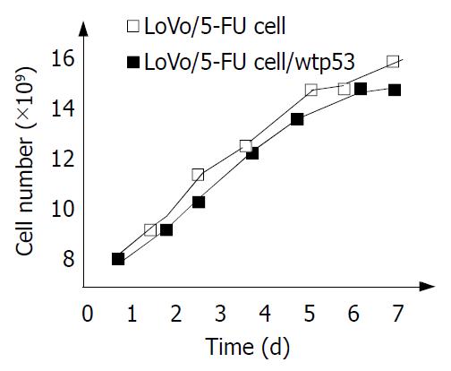
Figure 1 Effect of wild-type p53 on the growth rate of LoVo/ 5-FU cells.
Figure 2 Western blot analysis of wild-type p53 in LoVo/5-FU cell line and LoVo/5-FU cells transiently transfected with p53 expression vector (Ad-p53).
Lane 1: LoVo/5-FU cell line. Lanes 2, 3, 4, 5, 6: LoVo/5-FU cells transiently transfected with Ad-p53 for 2, 3, 4, 5, 6 d accordingly. Levels of wild-type p53 protein (lanes 2-6) were determined by Western-blot analysis with an antibody which could detect wild-type forms of p53 protein.
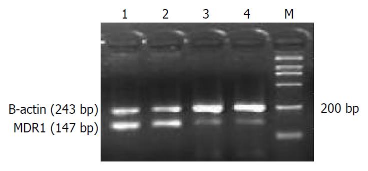
Figure 3 Results of RT-PCR for expression of MDR1 mRNAs in p53-null and wild-type p53 expressing LoVo/5-FU cells.
M: marker; Lanes 1 and 2: results of untransfected LoVo/5-FU cells; Lanes 3 and 4: results of transfected LoVo/5-FU cells.
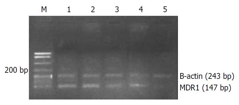
Figure 4 Results of RT-PCR for expression of MDR1 mRNAs of transfected LoVo/5-FU cells at different time.
M: marker; Lanes 1, 2, 3, 4, 5: the cells transfected with Ad-p53 for 2, 3, 4, 5, 6 d accordingly.
-
Citation: Yu ZW, Zhao P, Liu M, Dong XS, Tao J, Yao XQ, Yin XH, Li Y, Fu SB. Reversal of 5-flouroucial resistance by adenovirus-mediated transfer of wild-type
p53 gene in multidrug-resiatant human colon carcinoma LoVo/5-FU cells. World J Gastroenterol 2004; 10(13): 1979-1983 - URL: https://www.wjgnet.com/1007-9327/full/v10/i13/1979.htm
- DOI: https://dx.doi.org/10.3748/wjg.v10.i13.1979


